
Laser
Lasers are quite common in dentistry. Our clinic was one of the first in Auckland to purchase and start using dental lasers. At Marina Dentists, our clinicians are specialized in working with dental lasers. Lasers used in the dentistry field can be utilized for many different procedures relating to diseases of the teeth, gums, and oral mucosa, in surgical dentistry, implantology, and aesthetic dentistry, as well as during hygiene procedures. The advantages of the laser have already been seen by not only dentists but also by many patients. At Marina Dentists, we use lasers for the following procedures:
- Cutting the frenulum of the tongue, and upper and lower lip.
- Resection of the apex of the tooth root.
- Removal of the cysts.
Cutting the frenulum of the upper, lower lip, and tongue
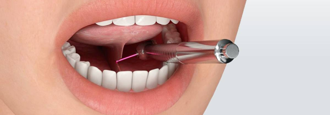 In the mouth, there are three thin films - the frenulum of both lips and the frenulum of the tongue. They play a huge role in the performance of the entire dental system. As an example, the lingual frenulum aids in various sound pronunciations, it is responsible for proper articulation and the ability to move the tongue. The frenulum of the lips interferes with the position of the front teeth, as well as the shape of the gums.
Why trim the frenulum
Depending on the frenulum structure, it can promote quite serious consequences to the patient. If the tongue frenulum is tight/short, the tongue will struggle to perform movements, which means that the patient cannot fully eat, and speech therapy problems appear. In infancy, the baby will have difficulty fully sucking on the mother's milk. Therefore, if this is the case, clinicians advise the frenulum to be trimmed in the first weeks or months of life. If trimming the frenulum of the tongue is performed in infancy, then problems with the short mucosa of the lips (usually the upper lip) may not be so obvious. Shortened and inelastic films can lead to gingival tension, reduction of its volume, and exposure of the roots of the front teeth. The consequences of exposure are increased sensitivity of the enamel, soreness, and active development of caries. While, too voluminous frenulum leads to overhanging of the mucosa over the surface of the teeth, which primarily affects the aesthetics of the smile and may lead to food entrapment.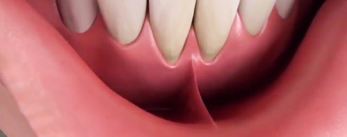 Thus, the indications for surgery are as follows:
- shortened frenulum,
- speech and pronunciation disorders of certain sounds,
- excessive tooth exposure,
- problems with nipple latching in infants,
- malocclusion caused by the position of the labial frenulum.
How does frenulum trimming work?
Trimming a baby's frenulum is quick and completely painless, taking only a couple of minutes. In adolescents and adults, the operation can take 20-30 minutes. Carrying out laser surgery in a dental clinic by the hands of professionals guarantees the absence of complications and minimizes the chances of surgical consequences.EXPERT OPINION
The possibility of using painkillers during frenulum cutting depends on the condition of the frenulum, the age of the patient, and his or her sensitivity. In children, the film is very thin, so the procedure takes only a few seconds, but in adult patients, anesthesia may be required, as the frenulum becomes tighter, and the operation can cause unpleasant and even painful sensations.
Alex Mechkov
Surgeon
Work experience 35+ years
Stages of the operation:
- injection of an anesthetic,
- dissection of the frenulum using a laser device, surgical instruments,
- suturing, which usually dissolves on their own 4-5 days after the operation.
ADDITIONAL TREATMENTS
After the operation of the tongue frenulum, speech therapy may be required. As it aids the child to speak and move the tongue again - this will also solve any pre-existing speech problems. If the shortened labial mucosa has caused aesthetic problems with the shape of the gums, an additional flap plastic surgery can be performed. Its purpose is the formation of a new gingival contour by dissection and displacement of the mucosa.After surgery
In most cases, there are no complications after trimming the frenulum of the upper, and lower lip or tongue, and the recovery takes only a few days. For a baby, it is best to feed him/her immediately, since the mother's milk acts as an anesthetic. While adult patients should temporarily avoid solid and viscous food. If there are problems with speech, then special classes with a speech therapist should be scheduled. There may be a slight discomfort to adult patients if stitches are placed, this will resolve after their resorption, which occurs usually after 4-5 days.Resection of the apex of the tooth root
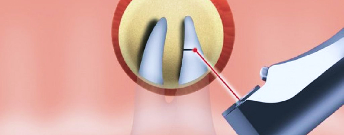 Resection of the apex of the root is a tooth-preserving procedure since it is actually the “last hope” for a tooth whose root has formed a cyst or granuloma. This operation involves the removal of the tumor along with a small part of the tooth root - in this way, the tooth itself is preserved and can even be used as the basis for fixing the prosthesis.
Indications
- cyst or granuloma more than one cm in diameter,
- tooth with a crown, canals - sealed,
- the presence of a pin in the tooth roots,
- root canals that are poorly sealed,
- therapeutic treatment of cysts or granulomas through the canals of the tooth is impossible.
Contraindications
- tooth mobility, when it is not necessary to save it,
- completely destroyed crown - in this case, a prosthesis cannot be fixed on the tooth,
- the size of the tumor is too large, the
- damage to the root of the tooth is large,
- general body problems: the cardiovascular system, infectious diseases, malignant tumors, diabetes mellitus,
- it is not recommended to use anesthesia.
Technology of the operation
The duration of resection of the apex of the tooth root is about an hour. It is necessarily carried out under anesthesia - even when drilling bone tissue, the patient is completely pain-free, and only slight discomfort is possible due to working with soft tissues.The stages of treatment are as follows
- diagnosis: the presence of a cyst or granuloma can often be determined only on x-rays since there are no symptoms. In the picture, the tumors look like blackouts near the tooth root,
- the removal of the nerve and the filling of the dental canals are carried out a couple of days before the operation, but not earlier since there is a high risk of purulent inflammation as a reaction to the filling. The nerve must be removed without fail since its inflammation and damage is possible,
- anaesthesia is a mandatory procedure before treatment since without anesthesia the operation is not possible,
- the gum is dissected, a small flap is exfoliated - the surgeon forms a cavity in the bone tissue through the hole by drilling
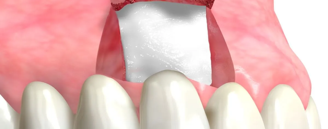
- through the hole in the gum and bone tissue, the doctor detects a tumor, carefully removes it together with the cut-off tip of the tooth root (sawed off with a drill),
- a part of the bone tissue extracted during the operation is replaced with an artificial material, which allows not to lose bone volumes. A protective membrane is applied on top, which dissolves on its own after a few weeks,
- the gum is sutured, and if necessary, drainage is applied between the seams to drain the pus - it is removed after 2-4 days.
Disadvantages of the operation
Resection of the apex of the tooth root is an operation that requires maximum attention from the patient and professionalism from the doctor. Among the shortcomings, one can single out the need for surgical intervention, as well as a rather long rehabilitation period, which is associated with taking medications and a number of restrictions (they relate to changes in lifestyle and diet).AFTER ROOT RESECTION EXPERT

After the operation, the patient must follow simple recommendations: stop eating for 2-3 hours, reduce physical activity for several days, and limit smoking. All hygiene procedures must be carried out carefully, without touching the wound.
Alex Mechkov
Surgeon
Experience 35+ years
Alternative solutions
Root resection is an operation that can significantly extend the life of single-rooted teeth near which a cyst or granuloma has formed. Alternatively, endodontic root canal treatment and drug therapy can be used if the tumor is small, as well as complete root removal in cases where the tumor is too large to cause significant tooth decay.Removal of a tooth
A tooth cyst is a tumor that forms in the area of the tooth roots. Most often, it occurs as a reaction to inflammation in order to prevent the further spread of the infection. A tooth cyst is a formation that is covered with a dense membrane. Treatment can be conservative or surgical (removal) - depending on the size of the tumor, its contents, and the degree of impact on the entire body.Diagnosis of a tooth
A tooth cyst, like a granuloma, practically does not manifest itself in any way - at the initial stages, the symptoms are completely absent. Only as the tumor grows, soreness, a lump on the gum, and an increase in body temperature occur. However, at regular preventive examinations, the doctor may suspect the presence of a tumor - with discomfort and soreness of previously treated teeth, it is they who fall into the “risk group”. The diagnosis is confirmed only by the results of an X-ray examination - in the picture, the cyst looks like a dark spot located around one or more tooth roots.Conservative treatment
Conservative or therapeutic treatment is used in the presence of a small cyst. Since the tumor is directly related to the tooth root, endodontic treatment is performed at the first stage - the dental nerve is removed, and the canals are thoroughly cleaned. A special medication is placed inside them, which promotes the resorption of the cyst, and a temporary filling is installed. At the same time, the patient is prescribed drug therapy. After 2-3 weeks, an X-ray examination is performed, which will show the condition of the cyst - during this time it should completely resolve. Gradually, its place will be filled with bone tissue cells. During the second visit, the temporary filling is removed, the root canals are sealed, and the top of the tooth is restored if necessary. This method, unfortunately, is not always effective - often it is not possible to completely get rid of pathological tissues and there is a high risk of recurrence of the disease. Therefore, after therapeutic treatment, regular monitoring of the condition of the tooth and periodontal tissues is required.Surgical removal of a tooth cyst: technologies
To date, surgical removal of a tumor is considered the most effective method of getting rid of the problem. And in 99% of cases, it is possible to save the tooth (upper, part of it), which later can even become support for attaching dentures. Any surgical intervention is performed under anesthesia, so the operation is painless. Unpleasant sensations occur only a few hours after it is carried out when the anesthetic ends. But they are associated exclusively with tissue injury.Tooth-preserving operations when removing a cyst
1. Cystectomy or resection of the root apex
The most popular, although one of the most difficult surgical operations is the treatment of a tooth cyst without removing it. It implies the removal of a cyst through a lateral incision in the gum and bone tissue, along with cutting off part of the tooth root - its top. The tooth nerve must first be removed and the canals are carefully sealed. Subsequently, such a tooth can last for several decades and even be used as a support for prosthetics.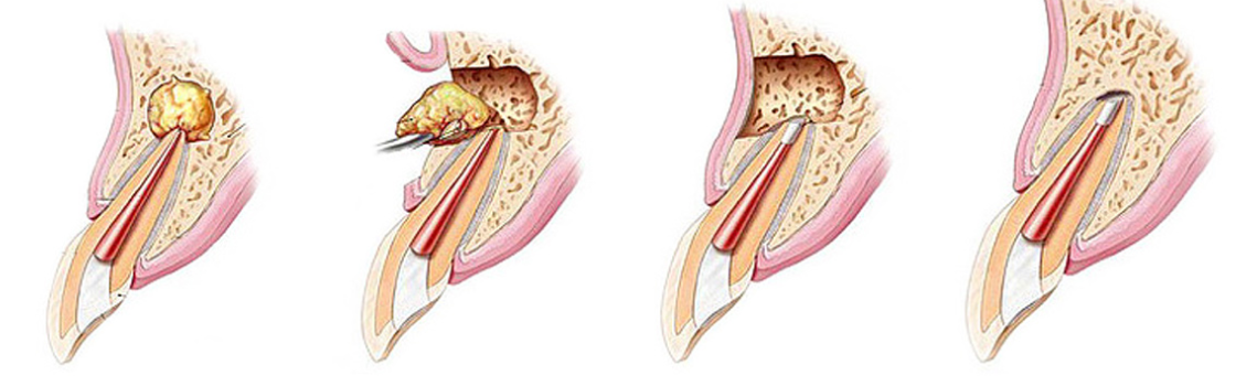
2. Hemisection
This operation involves the removal of a cyst along with one of the roots and the crown of a multi-rooted tooth. To begin with, the crown is sawn, then it is removed along with the root of the tooth. All pathological foci are removed - to provide additional access, the osseous-gingival flap can be peeled from the side. After the operation, the site of the tooth root is filled with synthetic material - artificial bone. The mucosa is sutured. A damaged tooth is necessarily restored by fixing a crown or bridge.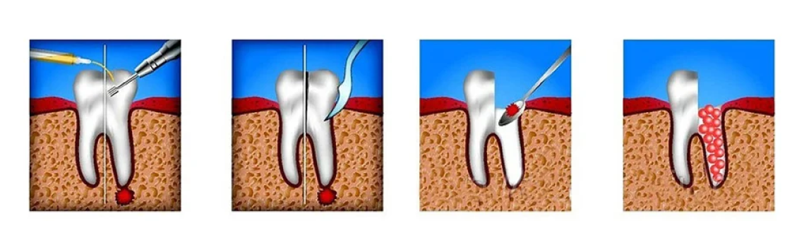
3. Cystotomy
This technology is used in the presence of a large tooth cyst when its complete removal can lead to thinning of the jawbone. The operation involves extracting fluid from the tumor by removing its front wall and creating a small hole in the bone tissue. Through it, the outflow of the internal contents of the cyst will be carried out. After its reduction in size, a complete extraction of the neoplasm is carried out.
In our clinic, an exclusively individual approach is practiced - after a thorough diagnosis, we select a treatment method that is suitable for a particular patient. At the same time, his general condition is necessarily taken into account so that surgical intervention does not harm his health.
Mechkov Alex
The surgeon with more than 35 years of practice
What to do if you had to extract a tooth
 Modern technologies allow us to remove cysts as carefully as possible for the teeth and at a reasonable price for Auckland. Subsequently, a single crown or bridge can be attached to them. However, if the tooth still has to be removed, the patient needs to think about prosthetics, and it is not worth delaying the restoration of the tooth - due to the lack of load, atrophy of the bone tissue will be observed, and adjacent teeth may shift towards the empty space, which will lead to the formation of gaps along the row.
Bridges or implant-supported single crowns. True, implantation will not work at the same time as the tooth is removed - in the presence of a voluminous cyst, it will take some time to restore the bone tissue, after which a dental implant can be installed.
Modern technologies allow us to remove cysts as carefully as possible for the teeth and at a reasonable price for Auckland. Subsequently, a single crown or bridge can be attached to them. However, if the tooth still has to be removed, the patient needs to think about prosthetics, and it is not worth delaying the restoration of the tooth - due to the lack of load, atrophy of the bone tissue will be observed, and adjacent teeth may shift towards the empty space, which will lead to the formation of gaps along the row.
Bridges or implant-supported single crowns. True, implantation will not work at the same time as the tooth is removed - in the presence of a voluminous cyst, it will take some time to restore the bone tissue, after which a dental implant can be installed.